Anti-Aging
Stem cell basis for skeletal aging
Aug
How aging contributes to bone loss is unclear. In aging mice, skeletal stem cells lose their ability to generate bone-forming cells called osteoblasts, and instead promote the generation of bone-resorbing cells called osteoclasts.
Aging is a key driver of bone-mass reductions and skeletal fragility. Insights into the identity of skeletal stem cells (SSCs) and other related progenitor cell populations that produce bone forming cells called osteoblasts have facilitated investigation into how aging affects skeletal cells.
Ambrosi et al. determined how with aging, the function of SSCs changes, contributing to bone loss and impaired skeletal regeneration. They isolated SSCs from the bones of young (2 month old) and aged (24 month old) mice and transplanted them into young recipient mice, in which they formed small masses of bone tissue.
This revealed 2 differences between young and aged SSCs. First, the bone mass produced by aged SSCs was much smaller and second, that aged SSCs exhibited an increased ability to promote the formation of osteoclasts, which are responsible for bone resorption (process by which osteoclasts break down the tissue in bones and release the minerals, such as calcium). Aging decreases the ability of SCCs to maintain a balance between bone formation and destruction.
They also surgically joined old and young mice, placing old and young SSCs in a shared blood circulation, which did relatively little overal to normalize bone formation in the aged mice. Therefore, the aging driven reprogramming of SSCs seems to be a function of the direct effects of aging and not being driven by aging associated factors circulating in the blood.
The researchers found that Colony stimulating factor 1 (CSF1), a soluble protein that promotes osteoclast maturation, is secreted at increased levels by aged SSCs. They mixed antibody molecules that bind and block the function of CSF1 together with BMP2 (protein with complex effects that can include promotion of bone formation) into a gel, then they placed around bone fractures in young and aged mice, which remarkably reversed the deficits in fracture healing seen in aged mice.
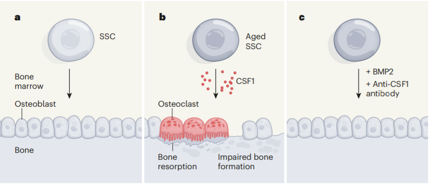
The first image below shows how SSCs serve as the cellular source of bone forming cells called osteoblasts. The second image represents the aging mice changes seen and how the SSCs become less able to generate osteoblasts and secrete increased levels of CSF1 which increases the generation of osteoclasts. The last picture represents a therapeutic strategy emerging from the model that they used, like antibodies that block CSF1, together with BMP2 to promote SCCs function.
Skeletal aging also encompasses a broader range of changes like the accumulation of senescent cells, increases in marrow fat content and changes in articular cartilage and bone architecture. More studies need to be performed to be able to completely understand the process of aging-associated changes in SSCs and the impact of other mechanisms of aging. The findings of this study can help in the development of new treatments that can target the changes seen here.
Source link: https://www.nature.com/articles/d41586-021-02118-0