Regenerative Medicine News and General Information
Can Mesenchymal Stem Cells Promote Antibacterial Defenses?
Mesenchymal stromal cells (MSCs) are adult multipotent progenitor cells that participate in the inflammatory, proliferative, and remodeling phases of tissue repair. The primary mechanism contributing to tissue repair occurs via paracrine signaling and, therefore, MSC-secreted factors are considered a promising therapeutic approach in regenerative medicine.
It has been demonstrated that MSC secretome, containing all secreted bioactive factors, stimulates dermal fibroblast migration and promotes angiogenesis in an equine model.
What is the MSC Secretome?
Paracrine stimulation is mediated by substances secreted by the MSCs as a response to the perceived environment. The secreted bioactive substances can be termed secretome.
Released factors include soluble proteins, free nucleic acids, lipids, and extracellular vesicles which can be further subdivided into apoptotic bodies, micro-particles, and exosomes.
The secretome from MSCs is believed to hold great potential for tissue repair and regenerative medicine.
Application of naïve MSC secretome in animal models has shown to significantly improve the pathology of various diseases such as graft versus host disease, autoimmune, and inflammatory diseases.
MSC and Wound Healing
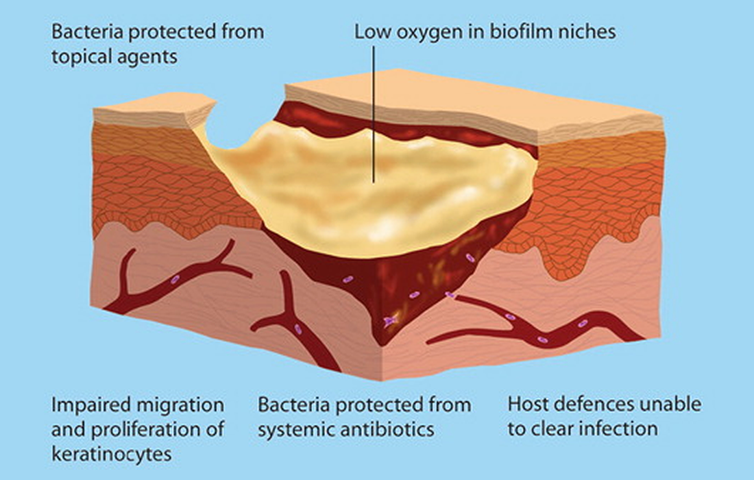
An important factor associated with compromised healing is wound colonization with pathogenic bacteria. In acute wounds, the main complication is infections, which, if not cleared, lead to wound chronicity. Infections can trigger an excessive inflammatory host response that suppresses the healing process. Subsequently, the skin barrier remains open, creating a vicious cycle by allowing continuous bacterial infestation. Bacterial wound colonization is a hallmark of chronic wounds, regardless of the underlying pathology.
Beside promoting skin tissue repair, researchers have found that MSC-secreted factors also possess antimicrobial properties, both against planktonic bacteria and bacteria in biofilms.
The skin is the most exposed organ of the body, it needs to guarantee a protective barrier against environmental insults such as microbes. The antimicrobial defense of the skin consists of complex interactions of resident skin and immune cells, as well as the skin microbiota. A healthy skin microbiota prevents invasion of the skin or wounds with pathogenic bacteria.
Study Found How MSCs Promote Antibacterial Defense in the Skin
A study published in the Journal STEM CELLS Translational Medicine tested the efficacy of the MSC secretome in a physiologically relevant ex vivo equine skin biofilm explant model and explored the impact of the MSC secretome on the antimicrobial defense mechanisms of skin cells.
The team found that secreted factors from equine MSCs significantly decreased viability of the bacteria methicillin-resistant Staphylococcus aureus in the skin models. They also found that MSCs secrete CCL2 that increases the antimicrobial activity of equine keratinocytes by stimulating the expression of antimicrobial peptides.
What is CCL2?
CCL2 also referred to as monocyte chemoattractant protein 1 (MCP1) and small inducible cytokine A2, is a small cytokine that recruits monocytes, memory T cells, and dendritic cells to the sites of inflammation produced by either tissue injury or infection.
Conclusions
The researchers found that MSCs secrete factors, such as the chemokine CCL2, that stimulate keratinocytes to (a) increase their expression of AMPs, such as ß-defensin and cathelicidin, and/or (b) enhance their efficacy of inhibiting bacterial growth, thus providing an indirect mechanism of the MSC secretome to reduce bacterial growth by stimulating innate antibacterial defense mechanisms in the skin.
They also found that MSC secretome stimulated the keratinocytes to increase their ß-defensin and cathelicidin expression, and that both have been shown to be effective against common skin pathogens such as S. aureus and Pseudomonas aeruginosa. Their results further support the value of MSC based treatments for infected wounds.
Source:
Charlotte Marx, et al. Mesenchymal stromal cell-secreted CCL2 promotes antibacterial defense mechanisms through increased antimicrobial peptide expression in keratinocytes. STEM CELLS Translational Medicine. 16 Sep 2021. https://doi.org/10.1002/sctm.21-0058
Carr MW, Roth SJ, Luther E, Rose SS, Springer TA (April 1994). “Monocyte chemoattractant protein 1 acts as a T-lymphocyte chemoattractant”. Proceedings of the National Academy of Sciences of the United States of America. 91 (9): 3652–6.
Xu LL, Warren MK, Rose WL, Gong W, Wang JM (September 1996). “Human recombinant monocyte chemotactic protein and other C-C chemokines bind and induce directional migration of dendritic cells in vitro”. Journal of Leukocyte Biology. 60 (3): 365–71.
Image from: https://www.magonlinelibrary.com/doi/abs/10.12968/bjon.2012.21.Sup20a.7

