Regenerative Medicine News and General Information
New Biomaterial Could Regenerate Intervertebral Disk Degeneration
Low back pain is a leading cause of disabilities throughout the world and engenders a tremendous socioeconomic burden. It has been estimated that intervertebral disk (IVD) degeneration accounts for 22–42% of patients with low back pain.
IVD degeneration frequently leads to more debilitating conditions, such as lumbar disk herniation, lumbar spinal stenosis, and spinal deformity, which are collectively called degenerative disk diseases.
There are different conservative treatments that may alleviate the symptoms but do not restore the degenerated IVD to its healthy state, and surgical interventions, such as decompression and fusion are palliative and do not resolve the pathology.
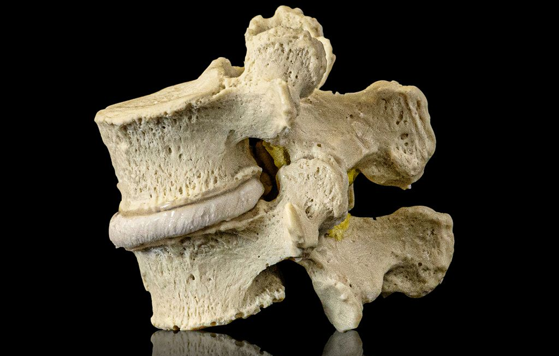
Anatomically, IVDs reside between vertebral bodies and constitute the spinal column. Functionally, IVDs resist the axial compression load of the spine and provide flexibility of the spinal column. This mechanical function is conferred by the anatomical structure of the IVD, in which nucleus pulposus (NP) in the center is surrounded by annulus fibrosus (AF) in the periphery. NP is viscoelastic and avascular and is composed of NP cells and extracellular matrix (ECM), which is produced and maintained by the NP cells. The ECM determines the mechanical properties of the NP.
The loss of nucleus pulposus (NP) precedes the intervertebral disk (IVD) degeneration that causes back pain.
To address this problem, a team of researchers at Osaka University and Kyoto University demonstrated that using cartilage tissue derived from human stem cells could help prevent the loss of functionality from IVD degeneration. The study appears in the journal Biomaterials.
Chondrocyte-like Cells
The team used induced pluripotent stem cells (iPSCs), which are reprogrammed somatic cells that have the same function as embryonic stem cells (ESCs). iPSCs are a promising cell source for regenerative therapy due to their unlimited proliferative and differentiative capacities.
These cells can be induced to become chondrocytes, which are cells that produce and maintain cartilage. The team developed iPSC-derived cartilaginous tissue (hiPS-Cart) for implantation into lab rats that had the nucleus pulposus (NP) removed.
The hiPS-Cart survived and occupied the nuclectomized space. Further scRNA-seq analysis revealed that hiPS-Cart cells changed their profile after implantation, differentiating into two lineages that are metabolically distinct from each other. However, post-implanted hiPS-Cart cells corresponded to chondrocyte-like NP cells only and did not develop into notochordal NP cells, suggesting that chondrocyte-like NP cells are nearly sufficient for NP function.
SOURCE:
Takashi Kamatani , Hiroki Hagizawa , Seido Yarimitsu , Miho Morioka , Saeko Koyamatsu , Michihiko Sugimoto , Joe Kodama , Junko Yamane , Hiroyuki Ishiguro , Shigeyuki Shichino , Kuniya Abe , Wataru Fujibuchi , Hiromichi Fujie , Takashi Kaito , Noriyuki Tsumaki (May , 2022). Human iPS cell-derived cartilaginous tissue spatially and functionally replaces nucleus pulposus. Elsevier. Retrieved from : https://www.sciencedirect.com/science/article/pii/S0142961222001302?via%3Dihub
IMAGE:
https://dynamicdiscdesigns.com/wp-content/uploads/2012/11/Lumbar-Spinal-Stenosis-Model-8.jpg

