Regenerative Medicine News and General Information
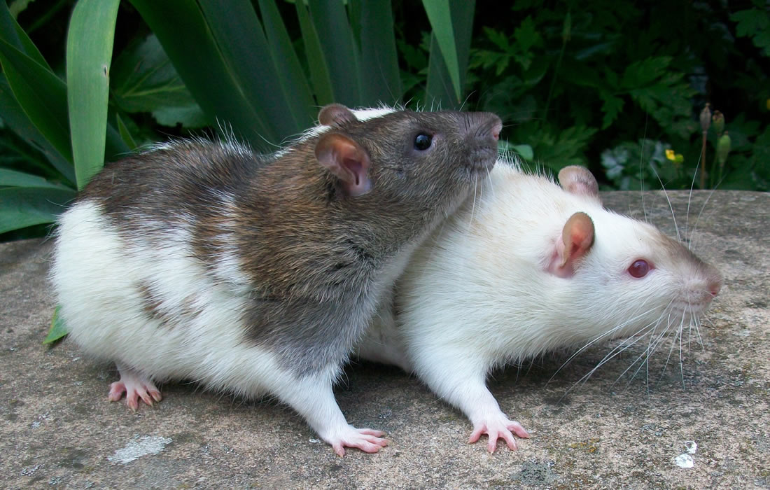
Immune System Mediates Volume and Function of Rat Brains
Researchers have established that biological sex plays a role in determining an individual’s risk of brain disorders.
For example, boys are more likely to be diagnosed with behavioral conditions like autism or attention deficit disorder, whereas women are more likely to suffer from anxiety disorders, depression, or migraines.
However, experts do not fully understand how sex contributes to brain development, particularly in the context of these diseases. They think, in part, it may have something to do with the differing sizes of certain brain regions.
University of Maryland School of Medicine researchers now believe they have identified the mechanism for why and how one brain region differs in size between males and females, according to a February study published in PNAS. The study conducted in rats found that immune system cells in the brains of females consume and digest neurons to sculp this brain region during development.
The researchers also found that tinkering with the size of this brain region, which forms in the first couple days of life, affected whether female rats still preferred the odor of male rats.
In rodents, this “odor preference” is an indicator of sexual partner preference with female rats typically preferring the odors of males. Although these rat inclinations do not directly apply to human sexual partner preferences, the findings demonstrate that changes to the brain that are determined by the immune system can later affect behavior.
Understanding in detail how biological sex and the immune system contribute to shaping the developing brain may one day help experts understand why certain brain diseases occur more likely in one sex versus another and could shed light on better ways to treat or prevent these conditions.
“Although there is much overlap between the brains of males and females, it seems to be the immune system that supplies much of the natural variation. This may occur because the immune system is designed for variability to be able to respond to a wide range of attacks from the outside world,” said UMSOM Dean Mark Gladwin, MD, Vice President for Medical Affairs at the University of Maryland, Baltimore, and the John Z. and Akiko K. Bowers Distinguished Professor.
For the current study, Dr. McCarthy and her colleagues examined a region located deep inside the brain that in male rats is two to four times larger than in female rats. This size difference also appears in the brains of people in a similar region, but the sex difference is not as pronounced.
When they closely examined different cell types in the male and female brain, they noticed that the immune cells in the female rat’s brains had formed more of the structures on their surface that immune cells use to eat other cells, called phagocytic cups. They also observed these immune cells digesting neurons. Typically, these immune cells eat debris, dead or dying cells, and cells infected with viruses or bacteria, rather than healthy brain cells.
When the researchers used a drug or an antibody to block the immune cells’ ability to eat neurons in rat brains, they found that this region in the female rat brains developed larger, similar to the size of the region in male rat brains.
The brain region analyzed in this study is known for controlling rat’s reproductive behaviors. For example, female rats typically prefer the odors of male rats when given a choice, and male rats prefer the odors of females.
The researchers found that females with the larger brain region due to their immune cells eating function being blocked no longer preferred the male rat odor and instead picked the female odor or had no preference at all.
“This finding adds to the evidence that the immune system plays a major role in determining certain sex differences in the brain that may ultimately lead to differences in the prevalence of developmental brain disorders,” said Dr. McCarthy. “Whether this process can be manipulated to develop new treatments for autism or anxiety remains to be seen, but it’s a promising avenue of research to explore.”
Sources:
Lindsay A. Pickett, Jonathan W. VanRyzin, Ashley E. Marquardt, Margaret M. McCarthy. Microglia phagocytosis mediates the volume and function of the rat sexually dimorphic nucleus of the preoptic area. Proceedings of the National Academy of Sciences, 2023; 120 (10) DOI: 10.1073/pnas.2212646120
University of Maryland School of Medicine. (2023, April 26). Immune system sculpts rat brains during development: Brain region size controls behavior preferences in adult rats. ScienceDaily. Retrieved April 28, 2023 from www.sciencedaily.com/releases/2023/04/230426210436.htm
Photo by Brendan Christopher from Pexels: https://www.pexels.com/photo/close-up-photo-of-mice-10398545/

