Regenerative Medicine News and General Information
Relationship Between Visceral Adiposity and Bone Density
Aug
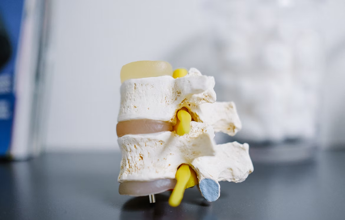
Osteoporosis is a systemic bone metabolic disease characterized by decreased bone mass, degeneration of bone tissue structure, which subsequently results in bone fragility and susceptibility to fractures.
The incidence of osteoporosis rises rapidly as the population ages, with approximately 1.5 million typical osteoporotic fractures (OF) of the spine, hip, and wrist occurring each year in the United States
The risk factors for osteoporosis are classified as uncontrollable and controllable factors, with the latter including low body weight, excessive drinking and smoking, and unhealthy lifestyles.
Recent studies have indicated that visceral fat accumulation is closely linked to osteoporosis. With the improvement of living standards and changes in dietary structure, the number of individuals with central obesity is increasing
The Visceral Adiposity Index (VAI), which reflects fat distribution and visceral fat metabolism, significantly, positively associated with lumbar bone mineral density (BMD), may help in the prediction, early screening, and evaluation of osteoporosis.
According to relevant mechanism studies, on the one hand, weight gain was commonly accompanied by increased fat mass, which inhibits the transformation of androgen into estrogen, decreases bone absorption and prevents bone mass loss. On the other hand the abnormal production of adipokines and activation of certain pro-inflammatory signaling pathways in obese patients could contribute to the production of TNF-α, IL-1, IL-6, and other inflammations, thereby stimulating osteoclast activity and increasing bone resorption.
Is there a relationship between VAI and BMD?
A study ,done by researchers at the Affiliated and the Second Affiliated Hospitals of Nanjing University of Chinese Medicine in China, included 9016 US residents included in the National Health and Nutrition Examination Survey (NHANES) during 2007-2010.
The researchers calculated a VAI for each subject using a sex-specific formula. The analysis included total femur, femoral neck, and lumbar spine bone mineral density measurements made by dual-energy-x-ray absorptiometry (DXA) bone scans.
VAI significantly, positively correlated with BMD after multiple adjustments for confounders. Every 1 unit increase in VAI is significantly associated with a 0.002 increase in lumbar BMD. The researchers also found similar significant, positive relationships between VAI and BMD for the total femur and for the femoral neck.
Participants in the highest VAI quartile had a 0.049g/cm2 higher BMD than those in the lowest VAI quartile, with the link remaining significant across different BMD quartile groups.
However, one age-related difference in the relationship emerged. Among teens and children in the data set ≤ 18 years old, among those with a VAI < 4.86, every 1 unit increase in VAI is associated with a significant 0.025g/cm2 higher lumbar BMD. But among those with a VAI > 4.86, a 1 unit increase in VAI is associated with a significant 0.027g/cm2 decrease in lumbar BMD.
There is a bidirectional relationship between this two variables , one in a high VAI may protect from osteoporosis because of the weight results to more work for the bones and with that a stronger bone structure, also with the increased conversion of the androgens to estrogens may protect the bone mass. But , on the other hand the pro-inflammatory status of obesity may increase bone resorption and decrease BMD. It seems to depend on the distribution of the fat tissue, visceral or subcutaneous.
SOURCE:
Shuai Chen, Guowei Zhou, Xiaohe Sun, Jie Jin, Zhiwei Li, (2022). Association between visceral adiposity index and bone mineral density: an NHANES study (pre-print document). Research square. Retrieved from: https://www.researchsquare.com/article/rs-1655251/v1
IMAGE:
Photo by Kenny Eliason on Usplash.