Regenerative Medicine News and General Information
Scientists Present Brain-Imaging Data for a Exosome Stroke Treatment that Supported Full Recovery
Therapeutic development for ischemic stroke has previously focused on small molecules, with anti-thrombotic, thrombolytic, and anti-inflammatory mechanisms of action. While approximately 4% of over 430 promising clinical trials for these small molecular therapeutics have reached world markets., ischemic stroke continues to remain a leading cause of death and long-term disability worldwide.
Identifying acute parameters which are predictive of long-term functional stroke outcomes has significant implications for characterizing patient injury severity, prognosis, and rehabilitation planning as well as offering improved efficacy assessments of neuroprotectants in preclinical studies
While identification of improved MRI biomarkers of stroke recovery and outcomes is essential, improving the ability to detect novel therapeutic options for stroke patients is warranted. Development and implementation of novel neuroprotective therapeutics are necessary to lead to holistic improvements in clinical patient outcomes and rehabilitation. Recent clinical trials have demonstrated the safety and efficacy of stem cell therapeutics in stroke patients with treatments leading to reduced lesion volumes and improved modified Rankin scale (mRS) and National Institutes of Health Stroke Scale (NIHSS) scores.
Exosomes are considered to be powerful mediators of long-distance cell-to-cell communication that can change the behavior of tumor and neighboring cells. The results of the study echo findings from other recent RBC studies using the same licensed exosome technology.
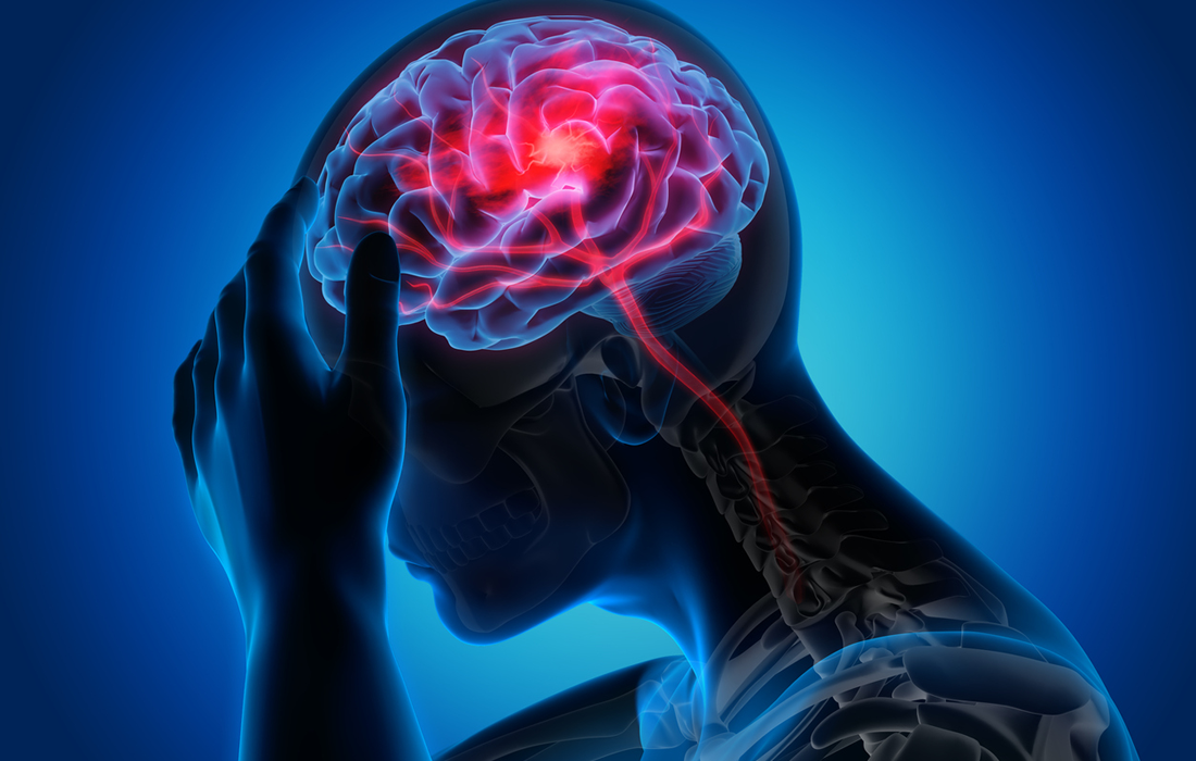
In a publication appearing this month in the journal Translational Stroke Research, animal scientists, funded by the National Institutes of Health, present brain-imaging data for a new stroke treatment that supported full recovery in swine, modeled with the same pattern of neurodegeneration as seen in humans with severe stroke.
Trauma from an acute stroke can happen quickly and can cause irreversible damage almost immediately. “Time is brain,” a phrase coined by stroke advocacy organizations in the late 1990s, captures the importance of acting on the first signs of stroke. In less than 60 seconds, warns the Stroke Awareness Foundation, an ischemic stroke kills 1.9 million brain cells.
In this observational study, the team analyzed brain images taken 24 hours after stroke. They then applied recovery scores, commonly used in human practice, based on swine gait, cadence, walking speed and stride length. By recording the relationship between brain measurements and functional outcomes, the new assessment scales can better help physicians predict how quickly a person will recover in real time.
Data from the team’s research showed that non-treated brain cells near the site of the stroke injury quickly starved from lack of oxygen and died — triggering a lethal action of damage signals throughout the brain network and potentially compromising millions of healthy cells.
However, in brain areas treated with exosomes that were taken directly from cold storage and administered intravenously, these cells were able to penetrate the brain and interrupt the process of cell death.
SOURCE:
Samantha E. Spellicy, Erin E. Kaiser, Michael M. Bowler, Brian J. Jurgielewicz, Robin L. Webb, Franklin D. West, Steven L. Stice.(December 6, 2019) Neural Stem Cell Extracellular Vesicles Disrupt Midline Shift Predictive Outcomes in Porcine Ischemic Stroke Model. Translational Stroke Research. Retrieved from : https://link.springer.com/article/10.1007/s12975-019-00753-4

