Regenerative Medicine News and General Information
Umbilical Cord Stem Cell Therapy Process
Sep
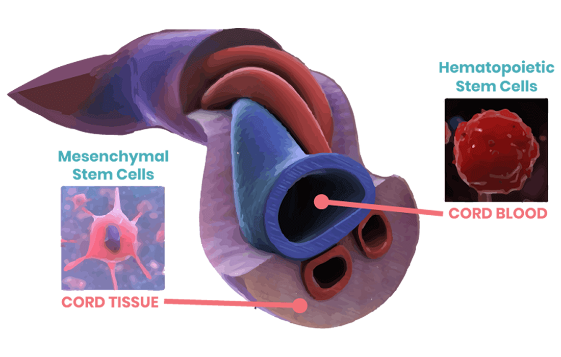
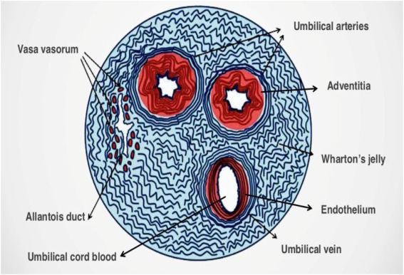
The umbilical cord is a noncontroversial source of mesenchymal stem cells. Mesenchymal cells are found in several tissue compartments of the umbilical cord, placenta and decidua. Umbilical cord mesenchymal stem cells derived from Wharton’s jelly (UCMSCs) have shown to have therapeutic potential, possibly as a substitute cell for bone marrow-derived mesenchymal stem cells for cellular therapy.
Mesenchymal stromal cells (MSCs), as defined by the International Society for Cellular Therapy, are plastic-adherent cells with a specific surface phenotype that have the capacity to self-renew and to differentiate into various lineages including bone, cartilage and adipose.
There are different steps in the in vitro culturing and storage of these cells. These steps are:
Isolation of Cells
The cells are obtained from full-term, normal C-section deliveries from consenting donors. These non-embryonic tissues undergo comprehensive medical, social and blood testing prior to procurement. After stringent screening the tissues are procured, processed and tested in accordance with standards established by the American Association of Tissue Banks (AATB) and FDA requirements.
- After delivery of the baby the umbilical cord is collected and stored in a sterile specimen cup containing 0.9% normal saline at 4°C until processing.
- The cord is handled in an aseptic fashion and processed.
- The length of the cord is estimated. The cord is manipulated in a sterile 10 cm petri dish and cut into 3-5 cm long pieces using a sterile blade.
- Blood vessels are removed from each piece after incising the cord lengthwise.
- The cord tissue is placed into a sterile 15 ml centrifuge tube and incubated in 3 ml of solution for 1 h at 37 °C.
- The tissue is moved to a new sterile dish filled with phosphate buffered saline and incubated for 30 minutes at 37 °C.
- After enzymatic digestion, the cord pieces are squeezed in a solution to remove as many cells from Wharton’s jelly as possible.
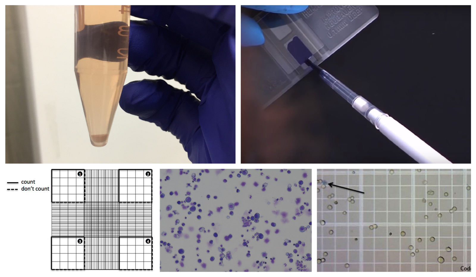
- The live cells are counted using a hemocytometer and plated in a 6-well tissue culture plate at a concentration of 30,000 cells per cm2 and incubated at 37 °C for 24-72 h.
- After that time, the cells are transferred to a new plate to allow cells to adhere.
- Cells in the original plate are fed with fresh medium and the cells are fed by the removal/replacement of half of the medium every 2-3 days till the cells reach approximately 80% confluence.
Passing the cells
- The cells are passaged when they are 80-90% confluent.
- The medium is aspirated off and cells are rinsed with sterile phosphate saline solution.
- The solution containing the cells is transferred to a 15 ml sterile centrifuge tube and centrifuged at 1000 rpm for 5 minutes.
- The cells are counted and transferred to a new plate or flask at a concentration of 10,000 cells per cm2 in fresh medium and then incubated at 37 °C.
Feeding the cells
- The cells are fed every other day or every 3 days.
- Half the medium in the plate is aspirated off and is replaced with fresh medium.
Counting the cells
- Cells are counted by trypan blue exclusion assay using a device known as hemocytometer.
Cryopreservation
- The cells collected for freezing should be in the growth phase.
- The cells in the freezing medium are transferred into a cryovial at 4 °C.
- The cryovial is transferred to a controlled rate cooler, maintained at 4°C and placed in the coldest part of the -80 °C freezer.
- The cryovial is transferred to a liquid nitrogen tank in a day or two.
- The vials are removed from the liquid nitrogen tank and plunged into a 37°C water bath with gentle swirling.
- Before the last ice crystal has melted, the vials are wiped down twice with 70% ethanol and moved to a biosafety cabinet.
- The cells are pelleted and resuspended in 1 ml of medium and a live/dead count is made.
Flowcytometry
- Tubes are incubated at room temperature in the dark for 15–20 min.
- After the incubation, the cells are washed with 2 ml of phosphate buffered saline.
- The cells are resuspended in 200 ml of phosphate buffered saline (Ca2þ free) and run through a flow cytometer.
- Typically for each tube, 10,000 events are collected and the data are analyzed using Cell Quest software.
At our clinic in Zignagenix we take every precaution to minimize potential risks of infectious disease testing and regenerative tests each step of its product along the way to ensure the highest quality safety standards are met and executed.
Source:
Kiran Seshareddy, et al. Method to Isolate Mesenchymal-Like Cells from Wharton’s Jelly of Umbilical Cord. Methods in Cell Biology, Academic Press,Volume 86,2008, Pages 101-119.