Regenerative Medicine News and General Information
Insights Into New Methods for the Early Detection and Better Monitoring of Multiple Sclerosis
Feb
Multiple sclerosis (MS) is a neurological disease that usually leads to permanent disabilities. One key feature of the disease is that it causes the patient’s own immune system to attack and destroy the myelin sheaths in the central nervous system. Myelin sheaths ensure that electrical impulses travel quickly and efficiently from nerve cell to nerve cell.
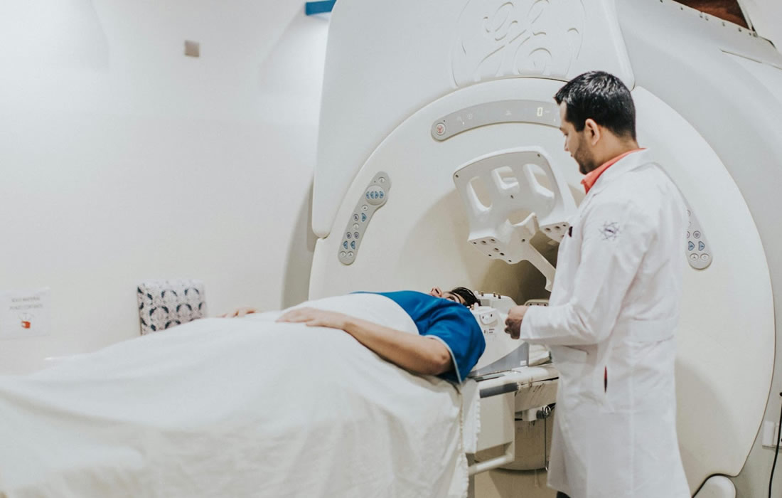
So far, however, it hasn’t been possible to visualize the myelin sheaths well enough to use this information for the diagnosis and monitoring of MS. Now researchers led by Markus Weiger and Emily Baadsvik, have developed a new magnetic resonance imaging (MRI) procedure that maps the condition of the myelin sheaths more accurately than was previously possible.
Conventional MRI devices capture only inaccurate, indirect images of the myelin sheaths.
That’s because most of these devices work by reacting to water molecules in the body that have been stimulated by radio waves in a strong magnetic field.
But the myelin sheaths, which wrap around the nerve fibers in several layers, consist mainly of fatty tissue and proteins.
Standard MRIs build their images primarily using the signals of the hydrogen atoms in this myelin water, rather than imaging the myelin sheaths directly.
The ETH researchers’ new MRI method solves this problem and measures the myelin content directly.
It puts numerical values on MRI images of the brain to show how much myelin is present in a particular area compared to other areas of the image.
Essentially, the darker the area and the smaller the number in the image, the more the myelin sheaths have been reduced.
This information ought to enable doctors to better assess the severity and progression of MS.
However, it is difficult to image the myelin sheaths directly.
That’s because the signals that the MRI triggers in the tissue are very short-lived; the signals that emanate from the myelin water last much longer.
To solve this problem, the researchers used a specially customized MRI head scanner.
This scanner is characterized by a particularly strong gradient in the magnetic field.
Generating such a strong gradient calls for a strong current and a sophisticated design.
As the researchers scan only the head, the magnetic field is more contained and concentrated than with conventional devices.
In addition, the system can quickly switch from transmitting radio waves to receiving signals; the researchers and their industry partners have developed a special circuit for this purpose.
The researchers have already successfully tested their MRI procedure on tissue samples from MS patients and on two healthy individuals. Next, they want to test it on MS patients themselves. Whether the new MRI head scanner will make its way into hospitals in the future now depends on the medical industry. “We’ve shown that our process works,” Weiger says. “Now it’s up to industry partners to implement it and bring it to market.”
Sources:
Emily Louise Baadsvik, Markus Weiger, Romain Froidevaux, Christoph Michael Schildknecht, Benjamin Victor Ineichen, Klaas Paul Pruessmann. Myelin bilayer mapping in the human brain in vivo. Magnetic Resonance in Medicine, 2024; DOI: 10.1002/mrm.29998
Materials provided by ETH Zurich. Original written by Christoph Elhardt. Note: Content may be edited for style and length.
ETH Zurich. “Visualising multiple sclerosis with a new MRI procedure.” ScienceDaily. ScienceDaily, 8 February 2024. <www.sciencedaily.com/releases/2024/02/240208122039.htm>.
Images from:
Photo by Charles Gonzhu
https://www.pexels.com/photo/doctor-examining-patient-on-mri-machine-15277954/